Quick answer
Retinal vein occlusion (RVO) is a blockage of one of the veins draining blood from the retina, causing haemorrhages and fluid build-up (macular oedema) that blur or reduce vision, usually in one eye and without pain. It most often affects people over 50 with high blood pressure, and is commonly treated with anti-VEGF injections.
What is Retinal Vein Occlusion?
Retinal vein occlusion (RVO) is the second most common retinal vascular disorder after diabetic retinopathy. It occurs when one of the veins draining blood from the retina becomes blocked, causing a back-up of blood and fluid that leads to retinal haemorrhages, macular oedema and, if untreated, permanent visual impairment. It most commonly affects people over 50, particularly those with hypertension, high cholesterol, or cardiovascular disease.
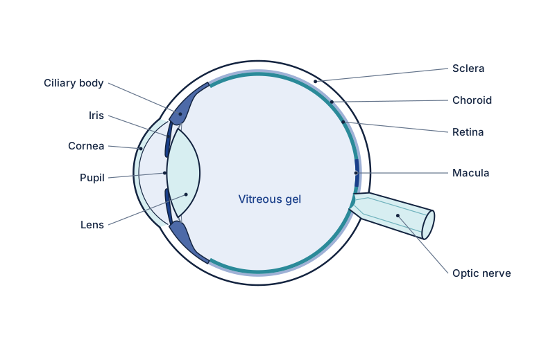
What are the symptoms of Retinal Vein Occlusion?
- Sudden painless loss or blurring of vision in one eye
- Distortion or grey area in central vision
- Sector-specific visual field loss (branch RVO)
- Floaters (from retinal haemorrhages)
What causes Retinal Vein Occlusion?
- Hypertension — the most important risk factor
- Hypercholesterolaemia
- Diabetes mellitus
- Glaucoma — raised intraocular pressure compresses the vein at the optic disc
- Thrombophilia (clotting disorders) in younger patients
How is Retinal Vein Occlusion diagnosed?
Dilated fundus examination, optical coherence tomography (OCT) to quantify macular oedema, fundus fluorescein angiography (FFA) to assess retinal perfusion. Systemic investigation to identify cardiovascular risk factors.
How is Retinal Vein Occlusion treated?
Intravitreal anti-VEGF injections (aflibercept, ranibizumab) are the first-line treatment for macular oedema associated with RVO, and have transformed visual outcomes. Intravitreal steroid implants (dexamethasone intravitreal implant) are an alternative, particularly for pseudophakic patients. Systemic cardiovascular risk factor modification is essential to reduce the risk of further vascular events.
How is this managed in Yorkshire?
This is not Mr Mohyudin’s operative list. He will not inject anti-VEGF for macular oedema from a vein occlusion. Use eye casualty or the medical-retina service; tell your GP about blood pressure and diabetes.
Frequently asked questions
What causes retinal vein occlusion?
The most important risk factor is high blood pressure. Others include raised cholesterol, diabetes, and glaucoma, which compresses the vein at the optic disc. In younger patients, clotting disorders may be responsible. Identifying and managing these cardiovascular risk factors is an important part of treatment.
Can vision recover after a retinal vein occlusion?
Vision often improves with treatment. Anti-VEGF injections are the first-line treatment for the macular swelling that causes vision loss in RVO and have transformed outcomes. The degree of recovery depends on how much the retina was affected and how promptly treatment begins.
How is retinal vein occlusion treated?
The main treatment for the associated macular oedema is intravitreal anti-VEGF injections; steroid implants are an alternative in selected cases. Alongside eye treatment, managing blood pressure, cholesterol and other cardiovascular risk factors is essential to reduce the risk of further vascular events.
Book a Consultation
Ask Spire Elland Hospital about current private consultation availability. A GP referral is not required for self-pay.
Book at Spire Elland ↗ Call 01422 324000Urgent — seek same-day care
Go to Eye Casualty (Huddersfield Royal Infirmary) or call NHS 111 now.
Related conditions
Self-Assessment Tool
Not sure if you need to be seen? Take our symptom checker.
Start self-assessment →