Quick answer
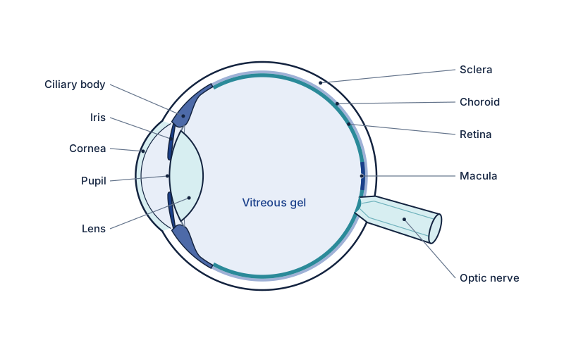
A macular hole is a small full-thickness break in the macula, the central, most sensitive part of the retina. It usually forms when the vitreous gel pulls on the macula during posterior vitreous detachment, causing blurred or distorted central vision. Most are idiopathic, occurring in people over 60, and more common in women.
What is Macular Hole?
A macular hole is a small break in the macula — the central, most sensitive part of the retina. It typically forms when the vitreous gel pulls on the macula as it separates from the retina (posterior vitreous detachment). Most macular holes are idiopathic, occurring in people over 60, and are more common in women. Left untreated, the hole tends to enlarge and vision deteriorates progressively. Surgery to close the hole achieves good results in the majority of patients.
What are the symptoms of Macular Hole?
- A blurred or missing spot in the centre of vision
- Distorted central vision — straight lines appearing wavy
- Difficulty reading fine print
- Reduced visual acuity, particularly for detailed tasks
What causes Macular Hole?
- Vitreous traction during posterior vitreous detachment (most common)
- High myopia (severe short-sightedness)
- Blunt eye trauma
- Epiretinal membrane contraction
How is Macular Hole diagnosed?
Optical coherence tomography (OCT) is diagnostic — it confirms the presence and stage of the hole, measures its size, and guides surgical planning. Visual acuity and Amsler grid assessment are also performed.
How is Macular Hole treated?
Small macular holes may occasionally close spontaneously, particularly in myopic patients. For full-thickness macular holes, vitrectomy surgery with internal limiting membrane (ILM) peeling and gas tamponade achieves closure rates of over 90%. Posturing (face-down positioning) for 5–10 days after surgery improves outcomes.
How is this managed in Yorkshire?
A full-thickness macular hole needs a vitreoretinal surgeon. Mr Mohyudin can diagnose on examination and OCT if available, then refer. Spire Elland cataract lists are not a substitute.
Frequently asked questions
What are the symptoms of a macular hole?
A macular hole typically causes a blurred or missing spot in the centre of vision and distorted central vision, where straight lines appear wavy. Patients often notice difficulty reading fine print and reduced visual acuity for detailed tasks. Peripheral vision is not affected, as the hole involves only the central macula.
How is a macular hole diagnosed?
Optical coherence tomography (OCT) is the diagnostic test. It confirms the presence and stage of the hole, measures its size and guides surgical planning. Visual acuity testing and Amsler grid assessment are also performed to evaluate central vision and detect the distortion that a macular hole produces.
How is a macular hole treated?
Small macular holes may occasionally close spontaneously, particularly in myopic patients. For full-thickness holes, vitrectomy surgery with internal limiting membrane peeling and gas tamponade achieves closure rates of over 90%. Face-down posturing for 5 to 10 days after surgery improves outcomes by helping the gas hold the hole closed.
Book a Consultation
Ask Spire Elland Hospital about current private consultation availability. A GP referral is not required for self-pay.
Book at Spire Elland ↗ Call 01422 324000Prompt assessment recommended
Related conditions
Self-Assessment Tool
Not sure if you need to be seen? Take our symptom checker.
Start self-assessment →