Quick answer
Keratoconus is a progressive eye condition in which the cornea (the clear front surface of the eye) gradually thins and bulges outwards into a cone-like shape. This causes progressive vision distortion that cannot be fully corrected with spectacles. It typically begins in the teenage years and may progress into the 30s and 40s.
What is Keratoconus?
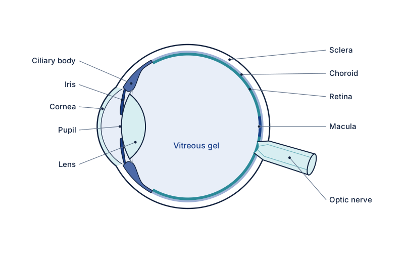
Keratoconus is a progressive eye condition in which the cornea — the clear front surface of the eye — gradually thins and bulges outwards into a cone-like shape. This irregular shape causes progressive distortion of vision that cannot be fully corrected with spectacles. It typically begins in the teenage years and may progress into the 30s and 40s, although the rate of progression varies considerably. Early diagnosis is essential to allow timely intervention to halt progression.
What are the symptoms of Keratoconus?
- Progressively worsening blurred vision, particularly at night
- Frequent changes in glasses prescription
- Increasing sensitivity to light and glare
- Ghost images or double vision in one eye
- Difficulty driving at night
- Contact lenses becoming increasingly uncomfortable or difficult to fit
What causes Keratoconus?
- Genetic predisposition — keratoconus runs in families in approximately 10% of cases
- Eye rubbing — a significant modifiable risk factor; patients should avoid all eye rubbing
- Atopic conditions (eczema, hay fever, asthma) are associated
- Connective tissue disorders (Marfan syndrome, Ehlers-Danlos syndrome)
How is Keratoconus diagnosed?
Corneal topography (mapping the shape of the corneal surface) and corneal tomography (measuring pachymetry and internal corneal structure) are essential. OCT and Scheimpflug imaging (e.g. Pentacam) provide detailed data on corneal curvature and thickness.
How is Keratoconus treated?
Spectacles and soft contact lenses are used in mild cases. Rigid gas permeable (RGP) or scleral contact lenses provide better visual correction as the condition progresses. Corneal cross-linking (CXL) — which stiffens the cornea using riboflavin and ultraviolet light — is the only proven treatment to halt progression and should be offered when progression is confirmed. Advanced cases may require corneal transplantation.
How is this managed in Yorkshire?
Mr Mohyudin can spot irregular astigmatism and refer. Rigid contact lenses and CXL belong with cornea and contact-lens colleagues in the region, not on an oculoplastic theatre list.
Frequently asked questions
Does corneal cross-linking improve vision?
Its main aim is to stop keratoconus getting worse rather than to improve vision. Most people still need glasses or contact lenses afterwards, but their cornea is more stable.
Who is corneal cross-linking for?
It is mainly for people with keratoconus that is still progressing, especially younger people, where strengthening the cornea early can help preserve future vision.
What are the early signs of keratoconus?
Early signs include progressively worsening blurred vision, particularly at night, and frequent changes in glasses prescription. Patients may notice increasing sensitivity to light and glare, ghost images or double vision in one eye, difficulty driving at night, and contact lenses becoming uncomfortable or hard to fit. Early diagnosis allows timely intervention.
Can keratoconus be stopped from getting worse?
Yes. Corneal cross-linking (CXL), which stiffens the cornea using riboflavin and ultraviolet light, is the only proven treatment to halt progression and should be offered when progression is confirmed. Avoiding all eye rubbing, a significant modifiable risk factor, is also important. Advanced cases may require corneal transplantation.
How is keratoconus diagnosed?
Keratoconus is diagnosed using corneal topography, which maps the shape of the corneal surface, and corneal tomography, which measures thickness and internal corneal structure. OCT and Scheimpflug imaging (such as Pentacam) provide detailed data on corneal curvature and thickness, allowing accurate assessment and timely intervention.
Book a Consultation
Ask Spire Elland Hospital about current private consultation availability. A GP referral is not required for self-pay.
Book at Spire Elland ↗ Call 01422 324000Prompt assessment recommended
Related conditions
Self-Assessment Tool
Not sure if you need to be seen? Take our symptom checker.
Start self-assessment →